
Fig. S2
- ID
- ZDB-FIG-110317-35
- Publication
- Yoo et al., 2010 - Differential Regulation of Protrusion and Polarity by PI(3)K during Neutrophil Motility in Live Zebrafish
- Other Figures
- All Figure Page
- Back to All Figure Page
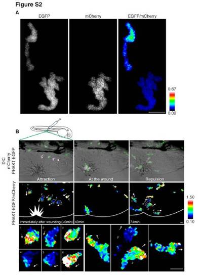
Ratiometric imaging (related to Figure 2) (A) Ratiometric image of EGFP/mCherry. The construct of mCherry was injected into embryos of Tg(MPO:GFP)uw as described in supplementary methods. There is no polarized signal in the bottom right cell expressing both constructs, although the top left cell expressing only EGFP shows high signals in the cell body due to volumetric effects. (B) Reversal of PI(3,4,5)P3-PI(3,4)P2 polarity (ratiometric imaging of PHAKT-EGFP/mCherry) when neutrophils leave the wound induced with a needle in the tail fin (Movie S2C). Tg(MPO:PHAKT-EGFP)uw and Tg(MPO:mCherry)uw were crossed to establish expression of both PHAKT-EGFP and mCherry in neutrophils efficiently. PI(3,4,5)P3-PI(3,4)P2 polarity is established towards the wound during attraction, and the polarity of PI(3,4,5)P3-PI(3,4)P2 is reversed to the opposite pole away from the wound during reverse migration from the wound. White arrows indicate direction of migration. Note high signals of PI(3,4,5)P3-PI(3,4)P2 at the leading edge regardless of the levels of background signals in the cytoplasm. Scale bars, 10 μm. |
Reprinted from Developmental Cell, 18(2), Yoo, S.K., Deng, Q., Cavnar, P.J., Wu, Y.I., Hahn, K.M., and Huttenlocher, A., Differential Regulation of Protrusion and Polarity by PI(3)K during Neutrophil Motility in Live Zebrafish, 226-236, Copyright (2010) with permission from Elsevier. Full text @ Dev. Cell