
Fig. 7
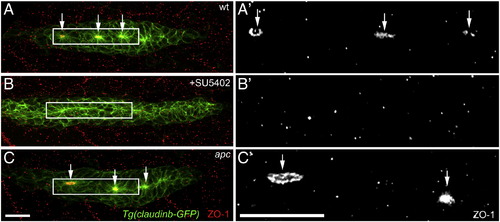
Epithelial rosette formation does not correlate with the proliferative behavior of cells. (A–C′) Labeling of the epithelial adherens junction protein ZO1 (red) in Tg(Claudinb-GFP) embryos. (A′–C′) ZO-1 labeling in the primordium. (A, A′) In wt primordia, epithelial rosette formation involves the apical constriction of cells within the primordium and the accumulation of apical adhesion complexes containing ZO-1 (arrows). Proliferation occurs in the unpatterned leading zone and immature rosettes. Cells in the most mature trailing rosette hardly proliferate. (B, B′) Inhibiting Fgf signaling using SU5402 causes a loss of apical constriction and ZO-1 accumulation in addition to a strong loss of proliferation. This finding argues that epithelial rosettes are not the cause of reduced proliferation in the trailing zone of wt primordia. (C, C′) in apc mutants, characterized by constitutively active Wnt/β-catenin and Fgf signaling in the primordium, apical constriction and ZO-1 accumulation occurs similarly as in wt primordia. In contrast to wt primordia, in apc mutants the trailing rosette is characterized by a high level of proliferation. Scale bars represent 20 μM. |
| Antibody: | |
|---|---|
| Fish: | |
| Condition: | |
| Anatomical Term: | |
| Stage Range: | 20-25 somites to Day 4 |
Reprinted from Developmental Biology, 349(2), Aman, A., Nguyen, M., and Piotrowski, T., Wnt/β-catenin dependent cell proliferation underlies segmented lateral line morphogenesis, 470-482, Copyright (2011) with permission from Elsevier. Full text @ Dev. Biol.