
Fig. 6
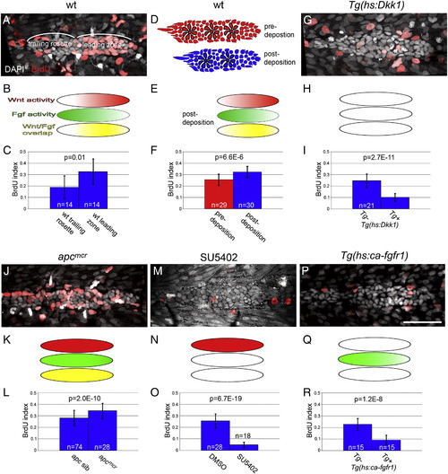
Wnt/β-catenin and Fgf signaling synergistically control proliferation in the primordium. (A,G,J,M,P) BrdU labeling in the primordium (red). Embryos were given a one hour pulse of BrdU at 32 hpf and immediately fixed. Nuclei are stained with DAPI (grey). (B,E,H,K,N,Q) Schematic representations of the distribution of Wnt/β-catenin signaling (red), Fgf signaling (green), and the region of overlap between these pathways (yellow). (C,F,I,L,O,R) Quantification of BrdU indexes for wt, Tg(hs:Dkk1), apc and SU5402 treated primordia. (A) Wt primordium with the leading zone and trailing rosette marked. Note the relatively low level of BrdU in the trailing rosette. (B) Schematic representation of Wnt/β-catenin and Fgf signaling activity in pre-deposition wt primordia. (C) Quantification of proliferation in the leading zone and trailing rosette of pre-deposition wt primordia. BrdU index is higher in the leading zone of wt primordia relative to the trailing rosette. (D) Schematic representation of a pre-deposition primordium harboring three rosettes (red) and a post-deposition primordium harboring two rosettes (blue). (E) Post-deposition primordia are shorter than pre-deposition primordial and have a smaller region free of wnt/β-catenin signaling. (F) Post-deposition primordial have a significantly higher BrdU index compared to pre-deposition primordia due to the loss of the relatively quiescent trailing rosette. (G) BrdU in a Tg(hs:Dkk1) primordium. Embryos were heatshocked six hours prior to BrdU incorporation. (H) Induction of Dkk1 leads to loss of Wnt/β-catenin and Fgf signaling in primordium. (I) Induction of Dkk1 significantly reduced BrdU. (J) apc mutant embryo. BrdU labeling is homogeneous throughout the mutant primordium. (K) apc mutation leads to the activation of both the Wnt/β-catenin and Fgf pathways throughout the primordium. (L) apc mutant primordial have a significantly higher BrdU index compared to wt sibs. The BrdU index in apc mutant primordia is similar to the index in the leading zone of wt primordia (compare (A) and (J)). (M) An embryo treated with 5 μM SU5402 from 22 to 32 hpf. (N) In SU5402 treated embryos Wnt/β-catenin signaling activity expands throughout the primordium. (O) The Fgf inhibitor SU5402 significantly reduces the BrdU index. (P) Induction of constitutively active Fgf receptor by heatshocking Tg(hs:ca-fgfr1) embryos reduces BrdU incorporation in the primordium. (Q) ca-fgfr1 expression leads to increased stimulation of Fgf signaling and a subsequent loss of Wnt/β-catenin signaling. (R) cafgfr1 expression leads to a significant reduction in BrdU incorporation. |
Reprinted from Developmental Biology, 349(2), Aman, A., Nguyen, M., and Piotrowski, T., Wnt/β-catenin dependent cell proliferation underlies segmented lateral line morphogenesis, 470-482, Copyright (2011) with permission from Elsevier. Full text @ Dev. Biol.