
FIGURE
Fig. 6
- ID
- ZDB-FIG-110221-25
- Publication
- Johnson et al., 2011 - mitfa is required at multiple stages of melanocyte differentiation but not to establish the melanocyte stem cell
- Other Figures
- All Figure Page
- Back to All Figure Page
Fig. 6
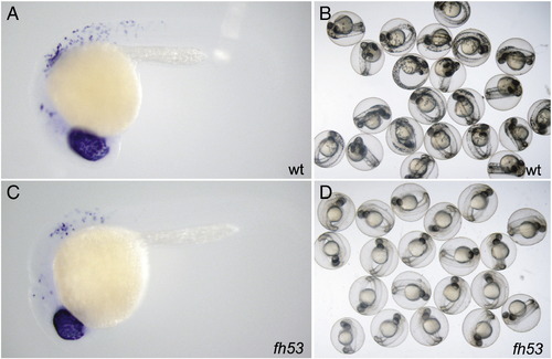
Embryos raised at permissive temperature to dct+ stage, when shifted show no melanized cells. A) Wild-type embryo showing dct expression in retinal pigment epithelium (RPE) and neural crest melanoblasts, B) wild-type embryos 20 h after upshift (stage equivalent to 45 h at standard temperature) showing differentiated melanocytes, C) mitfafh53 embryo showing dct expression at time of temperature upshift, and D) mitfafh53 embryos 20 h after upshift do not display any melanized cells save for RPE. In A and C, embryos were treated with 0.2 mM PTU to suppress melanin synthesis. |
Expression Data
| Gene: | |
|---|---|
| Fish: | |
| Condition: | |
| Anatomical Terms: | |
| Stage: | 26+ somites |
Expression Detail
Antibody Labeling
Phenotype Data
Phenotype Detail
Acknowledgments
This image is the copyrighted work of the attributed author or publisher, and
ZFIN has permission only to display this image to its users.
Additional permissions should be obtained from the applicable author or publisher of the image.
Reprinted from Developmental Biology, 350(2), Johnson, S.L., Nguyen, A.N., and Lister, J.A., mitfa is required at multiple stages of melanocyte differentiation but not to establish the melanocyte stem cell, 405-413, Copyright (2011) with permission from Elsevier. Full text @ Dev. Biol.