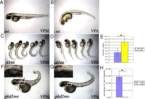
Inhibition of Class I HDACs can suppress the phenotypes of pkd2 mutants/morphants. (A and B) Treatment of 20 μm VPA from the shield stage leads to slight ventral curvature (B) on 2 dpf in wild-type embryos (wt) as compared to embryos treated with DMSO (A). (C–E) VPA can suppress the body curvature of pkd2/hi4166 mutant embryos. (C) shows mutant embryos treated with DMSO on 2 dpf. (D) shows mutant embryos treated with 20 μm VPA from 27 hpf on 2 dpf. (E) Average curvature angle in embryos treated with 20 μm VPM and 20 &,mu;m VPA. n = 5, *, P < 0.005, (F–H) Inhibition of kidney cyst formation in pkd2 morphants (pkd2mo) on 3 dpf by treatment of 20 μm VPA from the 20-somite stage. (F) shows a pkd2 morphants treated with 20 μm VPM. The arrow points to the cyst. (G) shows a morphant treated with VPA. White box: area shown in Insets. Insets show kidney cyst in F or the lack of cyst in G. (H) Average of percentage of embryos with kidney cysts from three independent experiments. Twenty-five to one hundred embryos were examined in each experiment. *, P < 0.005. 4166: embryos from pkd2/hi4166 heterozygous cross.
|
