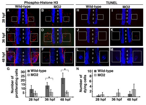
Pdlim7 knock-down pectoral fins have decreased cell proliferation. A-F: Dorsal view, anterior end of embryo is out of view to the bottom of image, of whole-mount anti-phospho-histone H3 (p-H3) antibody (red) staining on wild-type (A, C, E) and MO2 injected (B, D, F) embryos. Pectoral fins develop lateral to the 3rd somite, thus embryos were counterstained with MF20 (blue) to visualize somites, which are indicated by numbers (somite 1 refers to the most anterior somite). G: Quantification of p-H3 positive cells in pectoral fins of wild-type and MO2 injected embryos. 28 hpf wild-type n = 5, MO2 n = 5; p-value = 0.077. 36 hpf wild-type n = 4, MO2 n = 5; p-value = 0.036. 48 hpf wild-type n = 5, MO2 n = 5; p-value = 0.001. Experiment performed in triplicate, representative data from single replicate shown in G. Statistically significant p-values (<0.05) are denoted by asterisks. H-M: Whole-mount TUNEL assay on wild-type (H, J, L) and MO2 injected (I, K, M) embryos. Apoptotic cells in red with MF20 stained somites in blue, as described for p-H3 staining. N: Quantification of apoptotic cells in wild-type and MO2 injected embryos. 28 hpf wild-type n = 6, MO2 n = 4; p-value = 0.483. 36 hpf wild-type n = 6, MO2 n = 5; p-value = 0.05. 48 hpf wild-type n = 4, MO2 n = 6; p-value = 0.073. Experiment performed in triplicate, representative data from single replicate shown in N. White boxes indicate pectoral fin field at 28 hpf (A-B, H-I), 36 hpf (C-D, J-K), and 48 hpf (E-F, L-M).
|
