
- Title
-
Pdlim7 is required for maintenance of the mesenchymal/epidermal Fgf signaling feedback loop during zebrafish pectoral fin development
- Authors
- Camarata, T., Snyder, D., Schwend, T., Klosowiak, J., Holtrup, B., and Simon, H.G.
- Source
- Full text @ BMC Dev. Biol.
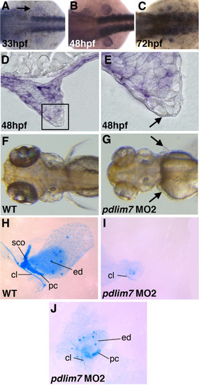
Pdlim7 is required for pectoral fin development. A-C: Whole-mount in situ hybridization using antisense RNA to show expression of pdlim7 in the developing pectoral fin mesenchyme at 33 hpf (A), 48 hpf (B), and 72 hpf (C). D-E: Sectioned embryos at 48 hpf of whole-mount in situ hybridization of pdlim7 show expression in fin mesenchyme. Boxed region in D is magnified in E to distinguish mesenchyme (purple color) and AER (arrow). F-G: Dorsal view of wild-type (F) and MO2 injected (G) embryos at 96 hpf. Arrows in G point to position of pectoral fin. H-J: Alcian blue stained cartilage preparations of dissected pectoral fins at 96 hpf. Wild-type (H), severe MO2 phenotype (I), and mild MO2 phenotype (J). I and J from same embryo. cl, cleithrum; pc, postcoracoid process; ed, endodermal disc; sco, scapulocoracoid. |
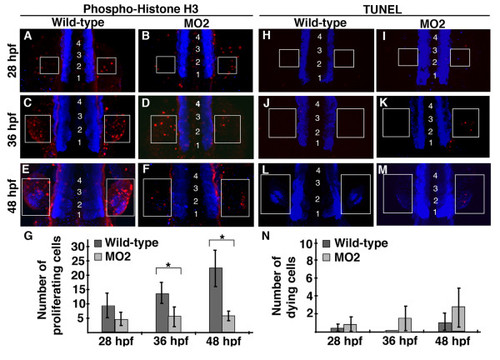
Pdlim7 knock-down pectoral fins have decreased cell proliferation. A-F: Dorsal view, anterior end of embryo is out of view to the bottom of image, of whole-mount anti-phospho-histone H3 (p-H3) antibody (red) staining on wild-type (A, C, E) and MO2 injected (B, D, F) embryos. Pectoral fins develop lateral to the 3rd somite, thus embryos were counterstained with MF20 (blue) to visualize somites, which are indicated by numbers (somite 1 refers to the most anterior somite). G: Quantification of p-H3 positive cells in pectoral fins of wild-type and MO2 injected embryos. 28 hpf wild-type n = 5, MO2 n = 5; p-value = 0.077. 36 hpf wild-type n = 4, MO2 n = 5; p-value = 0.036. 48 hpf wild-type n = 5, MO2 n = 5; p-value = 0.001. Experiment performed in triplicate, representative data from single replicate shown in G. Statistically significant p-values (<0.05) are denoted by asterisks. H-M: Whole-mount TUNEL assay on wild-type (H, J, L) and MO2 injected (I, K, M) embryos. Apoptotic cells in red with MF20 stained somites in blue, as described for p-H3 staining. N: Quantification of apoptotic cells in wild-type and MO2 injected embryos. 28 hpf wild-type n = 6, MO2 n = 4; p-value = 0.483. 36 hpf wild-type n = 6, MO2 n = 5; p-value = 0.05. 48 hpf wild-type n = 4, MO2 n = 6; p-value = 0.073. Experiment performed in triplicate, representative data from single replicate shown in N. White boxes indicate pectoral fin field at 28 hpf (A-B, H-I), 36 hpf (C-D, J-K), and 48 hpf (E-F, L-M). |
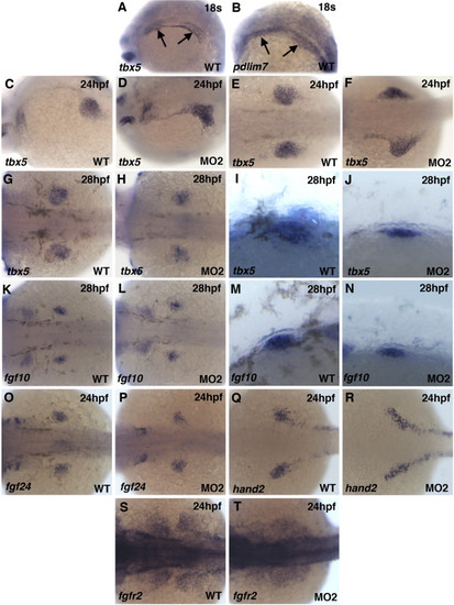
Migration and compaction defects in pdlim7 MO2 injected embryos. A-R: Whole-mount antisense RNA in situ hybridization of wild-type and MO2 injected embryos. tbx5 (A-H) expression at 24 hpf (A-D) and 28 hpf (E-H) in wild-type (A, C, E, G) and MO2 injected (B, D, F, H) embryos. G and H magnified lateral view of pectoral fin. fgf10 (I-L) expression at 28 hpf in wild-type (I, K) and MO2 injected (J, L) embryos. K and L magnified lateral view of pectoral fin. Expression at 24 hpf in wild-type and MO2 injected embryos of fgf24 (M-N), hand2 (O-P), and fgfr2 (Q-R), respectively. Head is positioned to the left. EXPRESSION / LABELING:
PHENOTYPE:
|
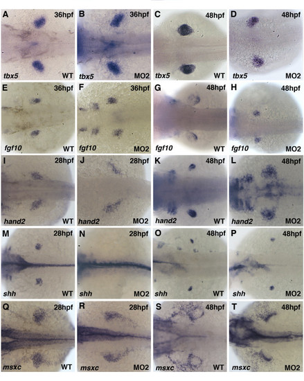
Pectoral fin mesenchymal gene expression in Pdlim7 knock-down embryos. Dorsal view of whole-mount antisense RNA in situ hybridization of wild-type (A, C, E, G, I, K, M, O, Q, S) and MO2 injected (B, D, F, H, J, L, N, P, R, T) embryos. A-D: tbx5 expression in wild-type and MO2 injected embryos at 36 hpf (A-B) and 48 hpf (C-D). E-H: fgf10 expression at 36 hpf (E-F) and 48 hpf (G-H). I-L: hand2 expression at 28 hpf (I-J) and 48 hpf (K-L). M-P: shh expression at 28 hpf (M-N) and 48 hpf (O-P). Q-T: msxc expression at 28 hpf (Q-R) and 48 hpf (S-T). Head is positioned to the left. |
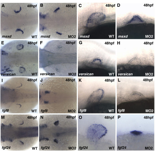
Disruption of AER gene expression in pdlim7 MO2 injected embryos. A-D: Expression of msxd at 48 hpf in wild-type (A, C) and MO2 injected (B, D) embryos. E-H: Expression of versican in wild-type (E, G) and MO2 injected (F, H) embryos. I-L: Expression of fgf8 in wild-type (I, K) and MO2 injected (J, L) embryos. M-P: Expression of fgf24 in wild-type (M, O) and MO2 injected (N, P) embryos. Dorsal views (A, B, E, F, I, J, M, N) and lateral views (C, D, G, H, K, L, O, P). Head is positioned to the left. |
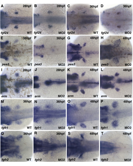
Fgf signaling pathway genes are disrupted after knock-down of Pdlim7. Dorsal view of whole-mount antisense RNA in situ hybridization of wild-type (A, C, E, G, I, K, M, O, Q, S) and MO2 injected (B, D, F, H, J, L, N, P, R, T) embryos. A-D: Expression of fgf24 at 28 hpf (A-B) and 36 hpf (C-D). E-H: Expression of pea3 at 36 hpf (E-F) and 48 hpf (G-H). I-L: Expression of erm at 28 hpf (I-J) and 48 hpf (K-L). M-P: Expression of fgfr1 at 36 hpf (M-N) and 48 hpf (O-P). Q-T: Expression of fgfr2 at 36 hpf (Q-R) and 48 hpf (S-T). Head is positioned to the left. |
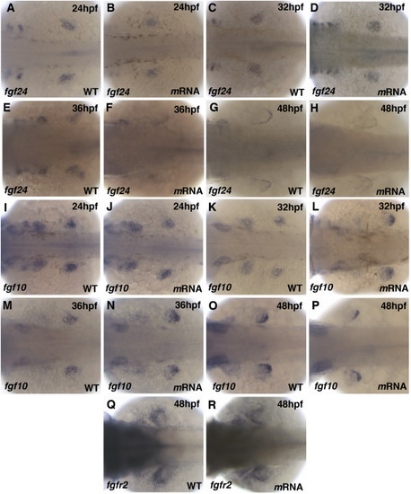
Pdlim7 overexpression does not alter Fgf signaling genes. Dorsal view of whole-mount antisense RNA in situ hybridization of wild-type (A, C, E, G, I, K, M, O, Q) and 100 pg synthetic pdlim7 mRNA injected (B, D, F, H, J, L, N, P, R) embryos. A-H: Expression of fgf24 at 24 hpf (A-B), 32 hpf (C-D), 36 hpf (E-F), and 48 hpf (G-H). I-P: Expression of fgf10 at 24 hpf (I-J), 32 hpf (K-L), 36 hpf (M-N), and 48 hpf (O-P). Q-R: Expression of fgfr2 at 48 hpf. |