|
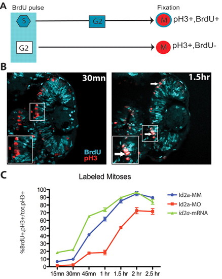
Id2a modulates the progression between S and M phase in proliferating retinoblasts. (A) Schematic of the percent labeled mitoses (PLM) paradigm. A 15-minute BrdU pulse marks S-phase retinoblasts, which then progress to M phase over time. Detection of M-phase cells with pH3 identifies those that were in S phase at the time of BrdU exposure. (B) Control retinas showing BrdU incorporation (cyan) and pH3 staining (red) at 30 minutes and 1.5 hours post-exposure. Many S-phase cells have progressed to M phase within 1.5 hours and are detected by BrdU/pH3 co-labeling (inset, arrow). (C) Graphical representation of PLM from 15 minutes to 2.5 hours post-exposure. Id2a-MO cells are delayed in progressing from S to M phase at all time points examined (n=4 retinas; **, P<0.003), whereas at 15 minutes, 30 minutes and 1 hours post-exposure, id2a-overexpressing retinoblasts have progressed from S to M phase more rapidly (n=4 retinas; **, P<0.025).
|