
Fig. 1
- ID
- ZDB-FIG-101011-2
- Publication
- Garavito-Aguilar et al., 2010 - Hand2 ensures an appropriate environment for cardiac fusion by limiting Fibronectin function
- Other Figures
- All Figure Page
- Back to All Figure Page
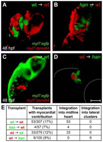
The role of hand2 in promoting cardiac fusion is not cell autonomous. (A-D) Confocal projections of mosaic hearts in live zebrafish embryos. (A-C) Lateral views, dorsal up. (D) Lateral view, dorsal down. (A,B) Wild-type host hearts expressing Tg(myl7:dsredt4) with integrated donor-derived cells expressing Tg(myl7:egfp) from wild-type (A) or han mutant (B) donors. Cells from wild-type or han mutant donors integrate indistinguishably into wild-type host hearts. (C,D) Wild-type (C) or han mutant (D) hosts expressing Tg(myl7:egfp) with integrated wild-type donor-derived cells expressing Tg(myl7:dsredt4). (D) In han mutant hosts, wild-type cells behave abnormally, remaining associated with lateral clusters of han mutant cardiomyocytes. Only the right-hand cluster is visible in this lateral view. Dotted lines indicate the embryo/yolk border (upper) and eye border (lower). Comparable clusters were observed in all nine chimeras examined, and no donor-derived cardiomyocytes were found outside of the clusters or at the midline. Scale bar: 100 μm. (E) Summary of results, indicating the ratio of hosts with donor-derived cardiomyocytes to all host embryos screened and the integration of donor-derived cardiomyocytes into the midline heart or lateral clusters. |