
FIGURE
Fig. 5
- ID
- ZDB-FIG-101007-16
- Publication
- Fang et al., 2010 - The expression patterns of minor fibrillar collagens during development in zebrafish
- Other Figures
- All Figure Page
- Back to All Figure Page
Fig. 5
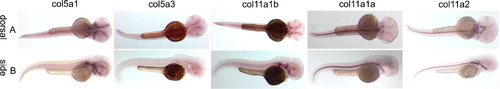
Spatial expression patterns of minor fibrillar collagen genes in zebrafish at 48 hpf by whole mount in situ hybridization. The expression patterns of Col5a1, Col5a3, Col11a1b, Col11a1a and Col11a2 are shown in both dorsal (A) and lateral (B) views. Expression is observed in the perioptic mesoderm, developing craniofacial cartilages, and presumptive retinal pigmented epithelium. Higher magnification views of patterns of expression are shown in Fig. 6 for 72 hpf. |
Expression Data
| Genes: | |
|---|---|
| Fish: | |
| Anatomical Terms: | |
| Stage: | Long-pec |
Expression Detail
Antibody Labeling
Phenotype Data
Phenotype Detail
Acknowledgments
This image is the copyrighted work of the attributed author or publisher, and
ZFIN has permission only to display this image to its users.
Additional permissions should be obtained from the applicable author or publisher of the image.
Gene expression patterns is full permission granted.
Reprinted from Gene expression patterns : GEP, 10(7-8), Fang, M., Adams, J.S., McMahan, B.L., Brown, R.J., and Oxford, J.T., The expression patterns of minor fibrillar collagens during development in zebrafish, 315-322, Copyright (2010) with permission from Elsevier. Full text @ Gene Expr. Patterns