|
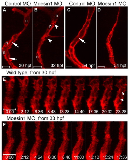
Moesin1 is required during ISVs tubulogenesis. (A-D) Confocal images of ISVs from Tg(flk1:mCherry-β-actin) living zebrafish embryos. (A,C) Embryos injected with 4 ng control MO. The lumen (arrows) is seen at 30 hpf (A) and is well formed at 54 hpf (C). (B,D) Embryos injected with 4 ng moesin1 MO. The primary lumen is not observed at 32 hpf (B), nor at 54 hpf (D), although a few putative vacuoles or intercellular spaces are observed (B, arrowheads). (E,F) Confocal time-lapse images of wild-type and Moesin1 knockdown Tg(flk1:mCherry-β-actin) embryos. The time format is minutes:seconds. (E) In wild-type embryos, formation of the primary lumen is observed (arrows) at 30 hpf. (F) In Moesin1 knockdown embryos, mCherry-β-Actin remains throughout the cytoplasm and the primary lumen is not observed at 33 hpf. n, nucleus.
|