
Fig. 8
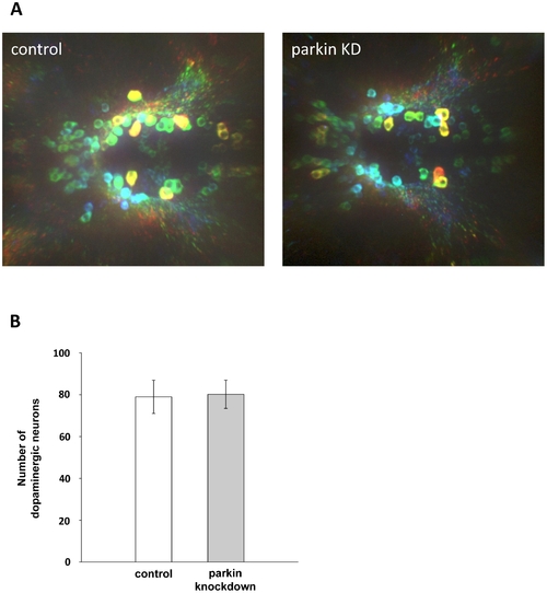
Parkin deficiency does not induce a loss of diencephalic dopaminergic neurons. (A) Whole mount immunostaining using an antibody for tyrosine hydroxylase (TH) as a marker for dopaminergic neurons. Staining of control GT-grip-injected and parkin GT-grip-injected zebrafish larvae shows no difference in the number of dopaminergic neurons. The neurons are depth-color-coded to illustrate the vertical position of the individual neurons in the brain and to allow the discrimination of neurons located on top of each other. Red neurons are located most dorsally, followed by yellow, green, blue and violet. Dorsal views of three-day-old larvae, anterior to the left. (B) Quantification of TH-positive dopaminergic neurons from 27 control and 27 parkin knockdown zebrafish larvae (parkin knockdown efficiency: 55%). TH-positive neurons were counted in a blinded manner, i.e. the researcher was blind to the knockdown status of the zebrafish larvae. |
| Gene: | |
|---|---|
| Fish: | |
| Anatomical Term: | |
| Stage: | Protruding-mouth |