
Fig. 1
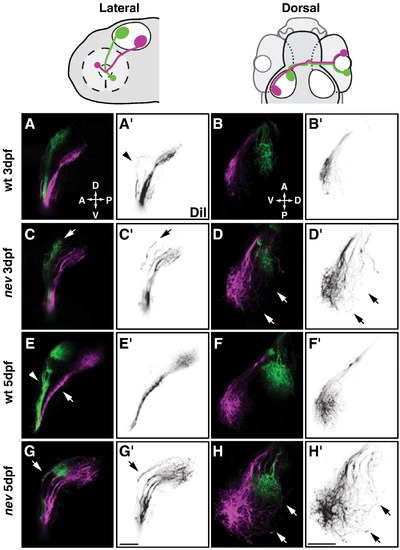
nevermind/cyfip2 is required for axon sorting and targeting of dorsonasal retinal axons. Confocal projections of dorsonasal axons (in magenta) and ventrotemporal axons (in green) in WT (A, B, E, F) and nev (C, D, G, H) at 3dpf (A–D) and 5dpf (E–H). (A′–H′) show only DiI injected dorsonasal axons in reverse contrast. Cartoons above show orientation of lateral views (A, C, E, G) and dorsal views (B, D, F, H). WT axons (A and E) from dorsonasal and ventrotemporal retina are topographically sorted in the ventral branch (arrow in E) and dorsal branch (arrowhead in E) of the optic tract, respectively. Arrowhead in A′ shows a few dorsal axons in WT projecting in the dorsal branch but turning before entering the tectum. Once on the tectum, WT axons project topographically to their target (B and F). However, in nev, dorsonasal axons are missorted in the dorsal branch of the optic tract (arrows in C and G), and project through the dorsal half of the optic tectum (arrows in D and H). Scale bars = 50 μm. |
| Fish: | |
|---|---|
| Observed In: | |
| Stage Range: | Protruding-mouth to Day 5 |
Reprinted from Developmental Biology, 344(2), Pittman, A.J., Gaynes, J.A., and Chien, C.B., nev (cyfip2) Is required for retinal lamination and axon guidance in the zebrafish retinotectal system, 784-794, Copyright (2010) with permission from Elsevier. Full text @ Dev. Biol.