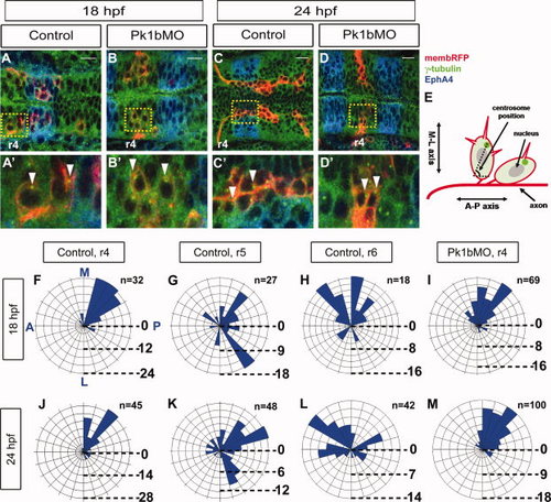
Centrosome remains primed for tangential migration in Pk1b-deficient neurons. A-D: Dorsal views of fixed embryos illustrating positioning of centrosome in migrating neurons. A: Control, 18 hpf. B: Pk1b morphant, 18 hpf. C: Control, 24 hpf. D: Pk1b morphant, 24 hpf. Red: zCREST1:membRFP transgene expression. Blue: EphA4 immunostaining, to label r3 and r5. Green: Υ-tubulin immunostaining, which allows visualization of centrosomes (bright puncta; white arrowheads) as well as the microtubule network (fainter green staining). Region of each cell with no green staining corresponds to nucleus. Flattened confocal z-stacks. Scale bars = 50 μm. A-D: Higher magnification views of regions denoted by yellow boxes in A-D. E: Schematic demonstrating measurement of centrosome position. A bisecting line was drawn from the centrosome (bright green puncta) through each neuron. The angle of this line, relative to the A-P axis was defined as the centrosome position. F-M: Rose diagrams illustrating the range of centrosome positions in migrating FBMNs. Numbers along right side indicate scale of y-axis (i.e., percentage of total neurons for each angle subset). n values indicated represent total number of neurons scored for each class. N = 5 embryos for each class.
|
