
Fig. 13
- ID
- ZDB-FIG-100616-104
- Publication
- Insinna et al., 2010 - Analysis of a zebrafish dync1h1 mutant reveals multiple functions for cytoplasmic dynein 1 during retinal photoreceptor development
- Other Figures
- All Figure Page
- Back to All Figure Page
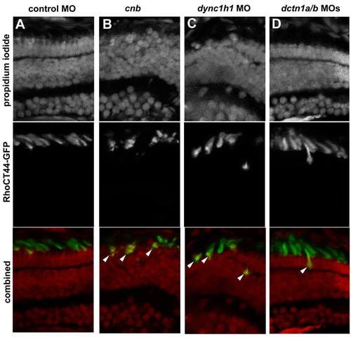
Efficient transport of Rhodopsin(CT44)-GFP to the outer segment is disrupted with loss of Dync1h1 or Dctn1. Cryosections of 3.5-dpf retina from transgenic Tg(1.3xops:xRhoCT44-GFP) embryos in which the carboxy-terminal 44 amino acids of Rhodopsin are fused with GFP. Localization of GFP was evaluated in photoreceptors from (A) control morphants, (B) cnb mutants, (C) dync1h1 morphants or (D) dctn1a/b morphants. Upper row shows propidium iodide staining to highlight nuclei. Middle row shows RhoCT44-GFP. Bottom row shows color combined images. Arrowheads indicate cells with mislocalized GFP to the cell body region. |
| Gene: | |
|---|---|
| Fish: | |
| Knockdown Reagents: | |
| Anatomical Terms: | |
| Stage: | Protruding-mouth |
| Fish: | |
|---|---|
| Knockdown Reagents: | |
| Observed In: | |
| Stage: | Protruding-mouth |