
FIGURE
Fig. 11
- ID
- ZDB-FIG-100616-102
- Publication
- Insinna et al., 2010 - Analysis of a zebrafish dync1h1 mutant reveals multiple functions for cytoplasmic dynein 1 during retinal photoreceptor development
- Other Figures
- All Figure Page
- Back to All Figure Page
Fig. 11
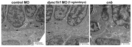
Ultrastructure of synaptic region in cnb mutants and dync1h1 morphants. TEMs of the photoreceptor synaptic region and outer plexiform layer of 3-dpf embryos. (A) Control morphants (3 ng/embryo) showed well developed synaptic input invaginations (asterisks) and numerous floating ribbons (arrows). (B) dync1h1 morphants (3 ng/embryo) had less defined synaptic input invaginations (asterisk) and significantly reduced number of floating ribbons (arrow). (C) cnb mutants did not show synaptic input invaginations and typically lacked ribbons. MO, morpholino. |
Expression Data
Expression Detail
Antibody Labeling
Phenotype Data
| Fish: | |
|---|---|
| Knockdown Reagent: | |
| Observed In: | |
| Stage: | Protruding-mouth |
Phenotype Detail
Acknowledgments
This image is the copyrighted work of the attributed author or publisher, and
ZFIN has permission only to display this image to its users.
Additional permissions should be obtained from the applicable author or publisher of the image.
Full text @ Neural Dev.