
Fig. 5
- ID
- ZDB-FIG-100520-23
- Publication
- Eames et al., 2010 - UDP xylose synthase 1 is required for morphogenesis and histogenesis of the craniofacial skeleton
- Other Figures
- All Figure Page
- Back to All Figure Page
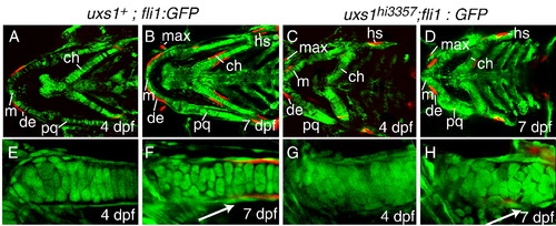
Cellular visualization of cartilage and bone morphologies in wild-type and uxs1 mutant larvae. (A–H) Optical sections of live Alizarin red-stained Tg(fli1:EGFP)y1 larvae, ventral views, at 4 dpf and the same individuals at 7 dpf. (E–H) Focus on the ceratohyal. In wild types (A, B, E, F), chondrocytes stacked and were lined with a flattened layer of perichondral cells (white arrow in F). Ossification centers stained with Alizarin red, reflecting perichondral bone formation in the ceratohyal and hyosymplectic and intramembranous ossification in the dentary and maxilla. In homozygous uxs1hi3357 animals (C, D, G, H); however, chondrocytes were disorganized, the perichondral sheath did not align properly (white arrow in H), and Alizarin red-positive ossification centers (dentary, maxilla, and ceratohyal) were severely reduced in perichondral and intramembranous sites. Abbreviations: ch, ceratohyal; de, dentary; hs, hyosymplectic; m, Meckel′s cartilage; max, maxilla; pq, palatoquadrate. |
| Fish: | |
|---|---|
| Observed In: | |
| Stage Range: | Day 4 to Days 7-13 |
Reprinted from Developmental Biology, 341(2), Eames, B.F., Singer, A., Smith, G.A., Wood, Z.A., Yan, Y.L., He, X., Polizzi, S.J., Catchen, J.M., Rodriguez-Mari, A., Linbo, T., Raible, D.W., and Postlethwait, J.H., UDP xylose synthase 1 is required for morphogenesis and histogenesis of the craniofacial skeleton, 400-415, Copyright (2010) with permission from Elsevier. Full text @ Dev. Biol.