
Fig. 2
- ID
- ZDB-FIG-100513-33
- Publication
- Lindsey et al., 2010 - From inflation to flotation: contribution of the swimbladder to whole-body density and swimming depth during development of the zebrafish (Danio rerio)
- Other Figures
- All Figure Page
- Back to All Figure Page
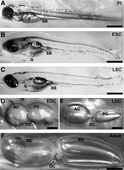
Photomicrographs of the stages of swimbladder development as viewed through the transparent body wall of larvae (A–C) or following excision from the coelomic cavity (D–F). All images show left lateral view. (A) Preinflation (PI) stage larva showing the location of the uninflated swimbladder (SB) in relation to the yolk sac (Y). (B) Early single-chamber (ESC) stage larva showing the inflated swimbladder adjacent to the developing gut (G). (C) Late single-chamber (LSC) stage larva showing the ovoid shape of the swimbladder in the coelomic cavity. (D) Early double-chamber (EDC) stage swimbladder displaying the anterior (AC) and posterior (PC) chambers connected by a wide ductus communicans (DC). (E) Late double-chamber (LDC) stage swimbladder showing the enlarged anterior chamber and constriction of the ductus communicans. (F) Adult swimbladder showing the ellipsoid-shaped anterior chamber and elongated posterior chamber. Scale bars: (A–C, F) 0.5 mm; (D, E) 0.2 mm. |