
Fig. 4
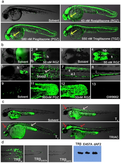
Ligand trap activities are drug responsive and reveal SNRM activities. (a) LT-PPARγ embryos show strong reporter activation in the presence of receptor specific agonist (uses as indicated). After heat pulse followed by overnight incubation with solvent or drug GFP expression in cells of the epidermis, CNS, eye, blood and heart (48 hpf; lateral view) is induced. Yellow arrow indicates GFP expression in heart. (b) Upper panel shows magnifications of embryo heads in ventral (b-1-2) and dorsal (b-3-4) orientation of 2 dpf fish treated with solvent or 50 nM Rosiglitazone (RGZ). B-2 shows reporter expression in eye (e), forebrain (fb), heart (h) and b-4 indicates GFP expression in hindbrain (hb), tectum (t) and anterior spinal cord (s). Middle panel shows drug responses in tissues of older embryos: Expression in heart (b-5; 5 dpf ventral view), in blood (b-6; 8 dpf), in the spinal cord (s) and endothelium layer ((el), b-7; 4 dpf, lateral view) and in the intestine ((I, b-8; 6 dpf). Embryos were subjected to a 30 minute heat pulse at 37°C, followed by incubation with 50 nM Rosiglitazone for 24 h. The lower panels show responses to: endogenous hormone in the tail epidermis of 3 dpf embryos treated with solvent (b-9); 500 nM Pioglitazone (b-11), 500 nM RGZ (b-12) and treatment with the antagonist GW9662 (500 nM). (c) TRβ LT embryos show tissue-specific responses to hormones and drugs. After 40 min heat induction, embryos (24 hpf) were incubated with compounds for 28 h in a 28°C incubator in the dark. Solvent (Ethanol) treated embryos show no GFP expression. T4 (2.5 μM) treated embryos show strong reporter activity in muscle and to a lesser amount in epidermis, brain and eye retina. T3 (2.5 μM) treatments result in similar responses but GFP induction in epidermis, brain and eye is stronger, and signal is also seen in blood cells. TRIAC (100 nM) treatments also induce GFP responses in brain, heart, eye, epidermis and muscle, and in addition, anterior spinal cord. Overlay pictures of bright field and GFP (75% transparent) are shown. Arrows indicate tissue-specific GFP responses: grey = muscle and red = brain. (d) TRβ responses in transgenic fish are dependent on a functional LBD. TRβwt, TRβE457A and TRβdAF2 embryos (24 hfp, F1) were heat pulsed and soaked in TRIAC (100 nM) as described in Figure 4c. The upper row shows strong (wt), weak (E457A) and no (dAF2) epidermal GFP expression in the tail. Western blot detected proteins show similar levels of TRβ transgene expression (right side). |