
Fig. 7
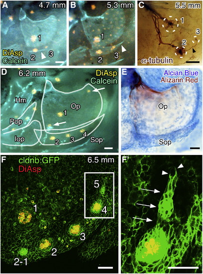
Development of the neuromast series on the subopercle. (A, B) Formation of OP3 along the extending subopercle visualized by staining with DiAsp and Calcein. Arrowhead indicates the subopercle. Body length of the larva is indicated in each panel. (C) Embryo immunostained for acetylated α-tubulin showing OP3 innervated by a nerve extending from OP2 (arrowhead). Neuromast positions are indicated by dotted circles. (D) Formation of OP4. Opercular bones are outlined. Hm, hyomandibular; Pop, preopercle; Iop, interopercle. Arrows indicate unidentified neuromasts in irregular positions on the opercle. (E) The same larva shown in D was stained with Alcian blue and Alizarin red. (F) Confocal image of a cldnb:gfp larva stained with DiAsp. (F′) Higher magnification of the boxed region in F, showing a budding structure (arrows). Arrowheads indicate the leading processes. Scale bars: 50 μm. |
| Gene: | |
|---|---|
| Fish: | |
| Anatomical Term: | |
| Stage: | Days 14-20 |
Reprinted from Developmental Biology, 340(2), Wada, H., Ghysen, A., Satou, C., Higashijima, S.I., Kawakami, K., Hamaguchi, S., and Sakaizumi, M., Dermal morphogenesis controls lateral line patterning during postembryonic development of teleost fish, 583-594, Copyright (2010) with permission from Elsevier. Full text @ Dev. Biol.