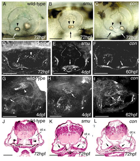
The ears of contf18b and smub641 homozygotes display a loss of posterior structures and a duplication of anterior structures. (A-C) DIC images of live ears, focussed at the level of the anterior otolith. Lateral views; anterior towards the left, dorsal towards the top. Both otoliths in con and smu ears (arrowheads, B,C) are small, lateral and ventral, resembling the anterior otolith (arrowhead, A) of wild-type embryos rather than the larger, medial posterior otolith (out of focus in A). Arrows indicate ventral sensory thickenings (maculae) underlying the otoliths. In the wild type, the anterior (utricular) macula lies under the anterior otolith on the ventral floor of the vesicle (A). In smu, a single ventral macula underlies the two small otoliths (B), while in con, a second ventral macula is found at the posterior of the ear (C). (D-I) Confocal images (projections of z-series) of ears stained with FITC-phalloidin to reveal the actin-rich stereocilia of sensory hair cells. (D-F) Lateral views; anterior towards the left, dorsal to top. (G-I) Dorsal views; anterior towards the left, medial towards the top. (D,G) Wild-type pattern. This is similar between 60 hpf and 4 dpf, but the number of hair cells increases in all patches during this time. Note the rounded anterior macula on the ventral floor of the vesicle (arrow) and the irregularly shaped posterior macula on the medial wall (arrowhead). Asterisks indicate the three cristae. In con and smu, the posterior macula is absent from the medial wall. In smu, a single ventral macula covers the ventral surface of the ear (arrow, E,H). In con, the anterior macula is present as normal (left arrow, F,I), but a second ventral macula is present at the posterior of the ear (right arrow, F,I). This resembles the posterior macula in shape but is smaller than normal. In a proportion of con and smu ears four cristae are present (E). (F,I) con ears with only two cristae, because of the relative immaturity of these ears. (J-L) Transverse paraffin sections (10 μm) through the otic vesicles, stained with Haematoxylin and Eosin. Dorsal towards the top. ot.v, otic vesicle; arrows, ventral maculae; arrowheads, posterior (medial) maculae; asterisks indicate cristae. Midline tissue is lost between the otic vesicles of smu and con embryos, so that the vesicles turn inwards towards the midline. However, all sensory patches are ventral; none are found on the medial wall. Scale bars: 50 μm.
|
