FIGURE

Fig. 6

ID
ZDB-FIG-100319-14
Publication
Gonzalez-Quevedo et al., 2010 - Neuronal Regulation of the Spatial Patterning of Neurogenesis
Other Figures
All Figure Page
Back to All Figure Page
Fig. 6

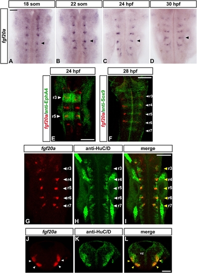
fgf20a Is Expressed in Neurons at Segment Centers
(A–D) Time course of fgf20a expression at 18 somites (A), 22 somites (B), 24 hpf (C), and 30 hpf (D). Embryos belong to the same batch and were developed for the same amount of time. Black arrowheads point at the center of r5. Scale bar, 50 μm. (E–I) Merge of confocal stacks of double-stained embryos. Scale bar, 100 μm. (E) In situ hybridization of fgf20a (red) and anti- EphA4 staining (green) to reveal r3 and r5 (white arrowheads) at 24 hr.
(F) In situ hybridization of fgf20a (red) and antibody staining for Sox9b (green) in 28 hpf embryo. White arrowheads indicate segment centers. (G–I) In situ hybridization of fgf20a (red) and antibody staining for the panneuronal marker HuC/D in 24 hpf embryos. White arrowheads indicate segment centers; yellow arrowheads show colocalization of fgf20a with specific HuC/D-expressing neurons.
(J–L) Double labeling of fgf20a (red) and HuC/D (green). Images show a merge of confocal stacks through r4 at 24 hpf in transverse sections (dorsal is to the top). In r4, fgf20a-expressing cells form clusters (white arrowheads) in the mantle zone and colocalize with specific HuC/D-expressing neurons (yellow arrowheads). VZ, ventricular zone. Scale bar, 50 μm. See also Figure S3.

Expression Data

Expression Detail
Antibody Labeling
Phenotype Data

Phenotype Detail
Acknowledgments
This image is the copyrighted work of the attributed author or publisher, and ZFIN has permission only to display this image to its users. Additional permissions should be obtained from the applicable author or publisher of the image.

Reprinted from Developmental Cell, 18(1), Gonzalez-Quevedo, R., Lee, Y., Poss, K.D., and Wilkinson, D.G., Neuronal Regulation of the Spatial Patterning of Neurogenesis, 136-147, Copyright (2010) with permission from Elsevier. Full text @ Dev. Cell