
Fig. 4
- ID
- ZDB-FIG-100211-4
- Publication
- Tay et al., 2010 - The iguana/DZIP1 protein is a novel component of the ciliogenic pathway essential for axonemal biogenesis
- Other Figures
- All Figure Page
- Back to All Figure Page
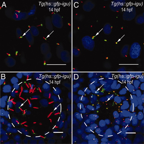
The Igu protein localizes to the bases of primary and motile cilia. A: Localization of green fluorescent protein (GFP) -Igu (green, arrows) to the bases of primary cilia (red) on paraxial mesodermal cells of a Tg(hs::gfp-igu) embryo. B: Localization of GFP-Igu (green, arrows) to the bases of Kupffer′s vesicle (KV) motile cilia (red) of a Tg(hs::gfp-igu) embryo. C: Localization of GFP-Igu (green, arrows) to basal bodies (red) of primary cilia on paraxial mesodermal cells of a Tg(hs::gfp-igu) embryo. D: Localization of GFP-Igu (green, arrows) to basal bodies (red) of KV motile cilia of a Tg(hs::gfp-igu) embryo. Embryos depicted in A and B were stained with anti-GFP (green) and anti-acetylated tubulin (red) antibodies, and those in C and D with anti-GFP (green) and anti-γ-tubulin antibodies (red). Nuclei were labeled with DAPI (blue). In B and D, circumference of KV is outlined with the dashed circle. A and C depict dorsal views of paraxial mesodermal cells; B and D represent ventral views of KV. Scale bars = 10 μm. |