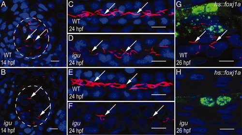
Motile ciliogenesis is less strongly perturbed in igu mutants. A: Motile cilia (arrows) in Kupffer′s vesicle (KV) of a wild-type embryo. B: Reduction in the numbers of motile cilia (arrows) in KV of an igu mutant embryo. C: Motile cilia (arrows) in the proximal segment of the pronephric duct of a wild-type embryo. D: Numbers of motile cilia (arrows) are reduced in the proximal pronephric duct of an igu mutant embryo. E: Motile cilia (arrows) in the distal segment of the pronephric duct of a wild-type embryo. F: Substantial reduction in the numbers of motile cilia (arrows) in the distal pronephric duct of an igu mutant embryo. G: A wild-type embryo with Foxj1a mis-expression (green) in muscle cells of the myotome, showing ectopic long cilia (arrows). H: No ectopic cilia formation in an igu mutant embryo with Foxj1a mis-expression (green) in myotomal muscle cells. In all panels, ciliary axonemes were labeled with anti-acetylated tubulin antibodies (red) and nuclei with DAPI (blue; 4′,6-diamidine-2-phenylidole-dihydrochloride). In A and B, circumference of KV is outlined with the dashed circle. Ectopic Foxj1a expression (green) in G and H was detected with antibodies against the HA tag at the C-terminus of the Foxj1a protein. A and B represent ventral views of KV; embryos in C-H are depicted anterior to the left, dorsal to the top. Scale bars = 10 μm.
|
