
FIGURE
Fig. 4
Fig. 4
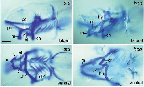
Photomicrographs of sturgeon (td204e) (A,B) and hoover (C,D) mutant larvae in which Meckel’s (first arch) is fused with the palatoquadrate. Lateral (A,C) and ventral (B,D) views of stained sturgeon and hoover larvae reveal that the ceratohyal is fused to the hyosymplectic as well. Additional unidentified pieces of cartilage occur in the vicinity of the basihyal and caudal to Meckel’s (A,B,D, arrows). In hoover mutant larvae the ceratohyal cartilage is sometimes divided into two pieces (D). Scale bar, 100 μm. |
Expression Data
Expression Detail
Antibody Labeling
Phenotype Data
| Fish: | |
|---|---|
| Observed In: | |
| Stage: | Day 5 |
Phenotype Detail
Acknowledgments
This image is the copyrighted work of the attributed author or publisher, and
ZFIN has permission only to display this image to its users.
Additional permissions should be obtained from the applicable author or publisher of the image.
Full text @ Development