|
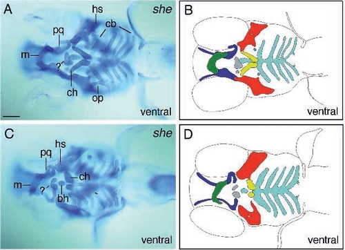
Photomicrographs (A,C) and diagrams (B,D) of ventral views of schmerle (th210) larvae, showing reductions of elements of the first two arches. B and D depict the elements of larvae shown in A and C. Larvae of one egg lay show variable reductions of elements (A,C). In the less-affected larva (A,B) the ceratohyal articulates with the hyosymplectic further to the anterior, but points posteriorly, whereas in the more severely affected specimens (C,D) these elements are reduced to two roundish structures. In both embryos additional pieces of cartilage are located in the vicinity of the basihyal indicated by (?) (C,A). Scale bar, 100 μm.
|