
Fig. 1
- ID
- ZDB-FIG-100128-12
- Publication
- Schütte et al., 2010 - Let It Flow: Morpholino Knockdown in Zebrafish Embryos Reveals a Pro-Angiogenic Effect of the Metalloprotease Meprin alpha(2)
- Other Figures
- All Figure Page
- Back to All Figure Page
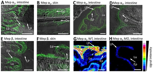
Distribution of meprin α1, α2 and β in zebrafish tissues. Immunofluorescence microscopy of cryosections from whole mount zebrafish, using specific peptide antibodies, revealed meprin α1 in the brush border cells of intestinal epithelia (Ep) (A) and epidermis (B), whereas meprin α2 was observed in the lamina propria mucosae (LP) only (C, D). Additionally, meprin β signals could be detected in the zebrafish intestine (Ep) and epidermis (Ed) (E, F respectively). To verify the efficiency of meprin knockdowns due to morpholino injection, we compared the fluorescence signal intensity in cryosections of wild type (G) and meprin α2 deficient embryos (H). Evidently, the expression of meprin α2 in the lamina propria (LP) of the intestine is largely decreased (G, H). Similar analyses of the meprin β morphant were not possible, due to the lethality within 24 hpf. (Ep: epithelium; Lu: lumen; LP: lamina propria; Ed: epidermis; De: dermis; scale bars: 25 μm; signal intensity was calculated with ImageJ V.1.41o). |
| Genes: | |
|---|---|
| Antibodies: | |
| Fish: | |
| Anatomical Terms: | |
| Stage: | Adult |