|
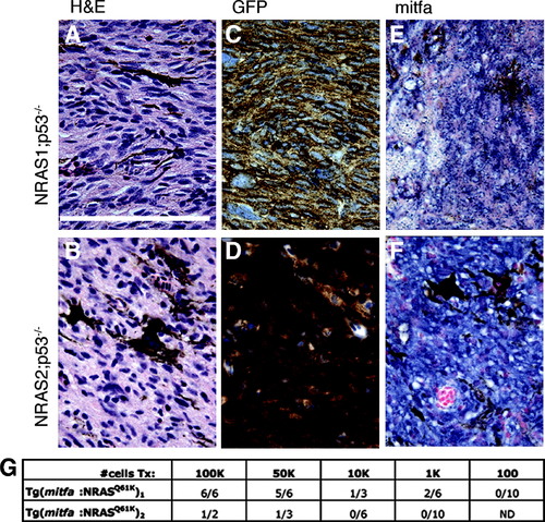
Histological examination of NRAS-driven tumors in zebrafish. (A, B) Hematoxylin and eosin (H&E) staining of melanomas reveals lesions with sporadic pigmentation and high levels of nuclear pleomorphism. (C, D) Standard immunohistochemistry against GFP revealed high levels of EGFP protein in these tumors, suggesting high and ubiquitous NRAS expression. (E, F) In situ hybridization to detect mitfa transcript indicates high levels of expression in these tumors. (G) Limiting dilution analysis of p53-null tumors from both transgenic lines transplanted intramuscularly into sublethally irradiated recipients. Scale bar = 10μm. (Color figure is available at liebertonline.com.)
|