
Fig. 4
- ID
- ZDB-FIG-100107-42
- Publication
- Christiansen et al., 2009 - Critical early roles for col27a1a and col27a1b in zebrafish notochord morphogenesis, vertebral mineralization and post-embryonic axial growth
- Other Figures
- All Figure Page
- Back to All Figure Page
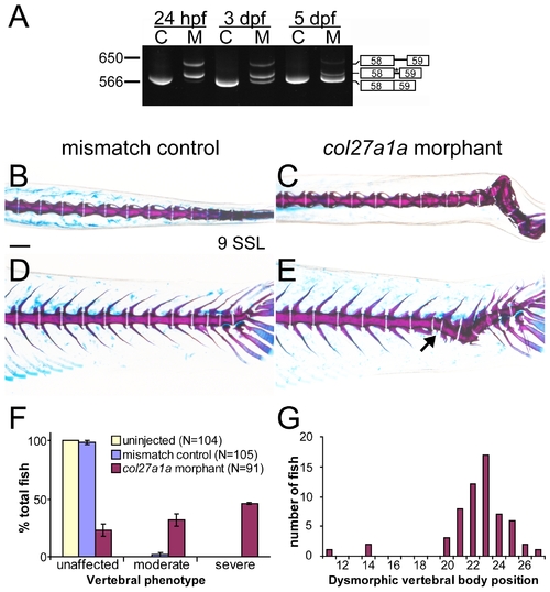
col27a1a morphants develop scoliosis and dysmorphic vertebrae. (A) Embryos injected with a SBMO targeting the exon 58 splice donor site of col27a1a showed abnormal splicing. Two abnormal splice forms including portions of intron 58 were generated. One splice form was predicted to generate a premature stop codon (asterisk). C, control; M, morphant. Dorsal and lateral views of col27a1a morphants (C, E) and mismatch control siblings (B, D) at ∼8.1–9.0 SSL (21 dpf) showed formation of scoliotic curves in the morphant vertebral column. Morphants were injected with a combined dose of 5 ng col27a1a SBMO+1 ng col27a1a TBMO. (F) By ∼8.1–9.0 SSL, 78% of col27a1a morphants had developed either moderate or severe vertebral defects. The mean from 3 clutches±standard error is shown. (E) Dysmorphic vertebrae without hemal or neural spines were also observed in the morphant vertebral column (black arrow). (G) These narrow, abnormal vertebrae were preferentially localized near the distal end of the tail. Vertebrae were counted beginning immediately distal to the 4 modified vertebrae of the Weberian apparatus. Scale bar in A represents 200 μm and applies to B–D. |
| Fish: | |
|---|---|
| Knockdown Reagents: | |
| Observed In: | |
| Stage: | Days 21-29 |