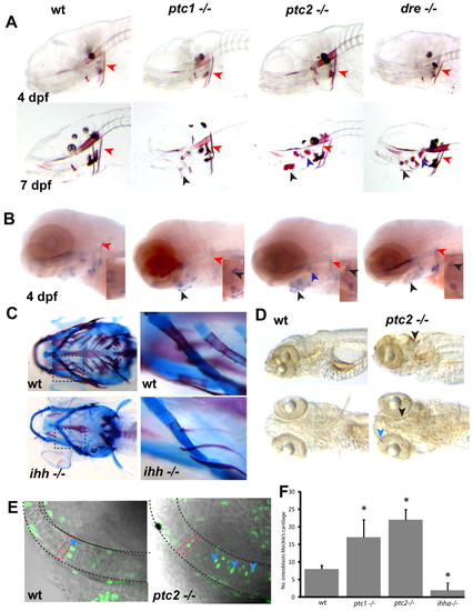
Levels of Hh signalling are critical for endochondral ossification and proliferation of chondrocytes. (A) Alizarin Red staining of representative Hh mutants reveals no differences at 4 dpf. At 7 dpf, however, premature mineralisation of endochondral bone elements can be seen (black arrowheads mark the ceratohyal, purple arrowheads mark the hyosymplectic), while the extent of ossification in the cleithrum is unchanged (red arrowheads). (B) In situ analysis of mutants at 4 dpf reveals increased expression of osterix in the endochondral bone, where premature mineralisation is later seen (black arrowheads point to ceratohyal, purple to hyosymplectic), while osterix expression is unchanged or slightly reduced in dermal bone elements such as the cleithrum (red arrowheads). Insets show ventral views of osterix expression in the ceratohyal. (C) Alcian Blue/Alazarin Red double staining of 17 dpf ihha and wild-type sibling fish. The second panel is an enlargement of the boxed area for each genotype. (D) Whole-mount antibody staining for BrdU incorporation between 4.5 and 5 dpf in wild type and ptc2 mutants. Excessive proliferation can be seen in the ptc2 mutant in the tectum (black arrowhead) and jaw cartilages (blue arrowhead). (E) Representative single confocal images of the Meckel′s cartilage in 5 dpf zebrafish. Proliferating chondrocytes (blue arrowheads) are labelled with anti-BrdU (green), the dashed red line shows the shape of a single chondrocyte. (F) Quantitation of BrdU-positive cells in the Meckel′s cartilage at 5 dpf. Data are mean±s.d. taken of at least five fish per genotype. *P<0.01 versus wild type. wt, wild type.
|
