
Fig. 1
- ID
- ZDB-FIG-091116-14
- Publication
- Mullins et al., 1996 - Genes establishing dorsoventral pattern formation in the zebrafish embryo: the ventral specifying genes
- Other Figures
- All Figure Page
- Back to All Figure Page
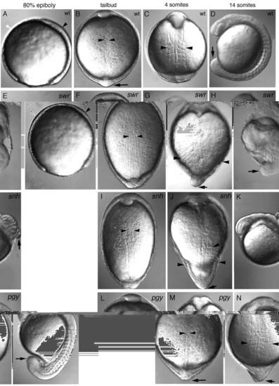
Morphological defects visible in live mutant gastrulae of swr, snh and pgy. Dorsal is to the right in lateral views and animal pole is up in dorsal views. (A-D) Wild type, (E-H) swr (class 5 phenotype), (I-K) snh (class 4 phenotype), (L-N) pg (class 3 phenotype). A lateral view of an 80% epiboly stage wild-type (A) and swr mutant (E) embryo showing the thickened ventral side (white arrowhead) and thinner dorsal axis (black arrowhead) in swr. A dorsal view at bud stage of wild-type (B), swr (F), snh (I), and pgy (L) embryos (the notochord is delineated by arrowheads). Dorsal views of 4- somite stage embryos: wild-type (C), swr (G), snh (J), and pgy (M). The most lateral extent of the somites is marked by arrowheads; an arrow indicates the position of the tailbud, which is not visible in the wild type (C) because it has extended around the yolk. (D,H,K,N) Lateral views of 14-somite stage embryos: wild-type (D), swr (H), snh (K), and pgy (N) (the tailbud is indicated with an arrow). |
| Fish: | |
|---|---|
| Observed In: | |
| Stage Range: | 75%-epiboly to 14-19 somites |