
|
Fig. 1
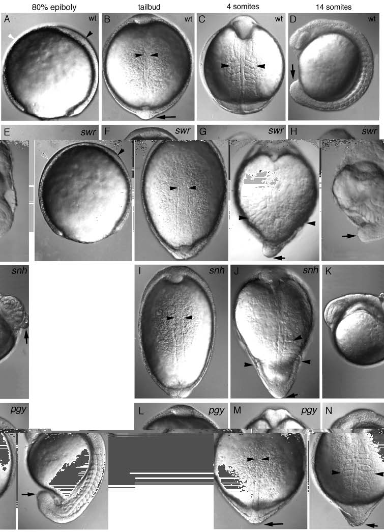
Morphological defects visible in live mutant gastrulae of swr, snh and pgy. Dorsal is to the right in lateral views and animal pole is up in dorsal views. (A-D) Wild type, (E-H) swr (class 5 phenotype), (I-K) snh (class 4 phenotype), (L-N) pg (class 3 phenotype). A lateral view of an 80% epiboly stage wild-type (A) and swr mutant (E) embryo showing the thickened ventral side (white arrowhead) and thinner dorsal axis (black arrowhead) in swr. A dorsal view at bud stage of wild-type (B), swr (F), snh (I), and pgy (L) embryos (the notochord is delineated by arrowheads). Dorsal views of 4- somite stage embryos: wild-type (C), swr (G), snh (J), and pgy (M). The most lateral extent of the somites is marked by arrowheads; an arrow indicates the position of the tailbud, which is not visible in the wild type (C) because it has extended around the yolk. (D,H,K,N) Lateral views of 14-somite stage embryos: wild-type (D), swr (H), snh (K), and pgy (N) (the tailbud is indicated with an arrow).