
Fig. S3
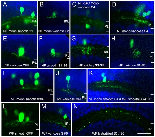
Morphology of additional types of amacrine cells in Ptf1a:GFP DNA-injected embryos (120 hpf). Micrographs show single images only and some of the joining neurites and/or somas are thus not in focus in the shown images. The nuclear stain DAPI was used to reveal the retinal layers. As described in Figure 7, different subtypes can be distinguished by the stratification depth, breadth, neurite arbor width and smooth or beaded neurite morphology. (A-H, K) Narrow-field amacrine cell types. (I, J) Medium-field amacrine cell types. (K-N) Wide-field amacrine cell types. dAC, displaced amacrine cell; IPL, inner plexiform layer; MF, medium-field; mono, monostratified; NF, narrow-field; WF, wide-field. Scale bar = 20 μm. |