
FIGURE
Fig. 6
Fig. 6
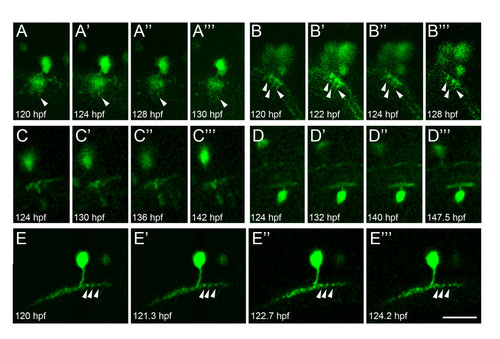
Ptf1a:GFP amacrine cells at 120 hpf have stable neurite arbors. Single time-lapse images of different amacrine cells imaged for up to 24 hours. The GFP signal is relatively weak in some cells but, nonetheless, the stratification depth extent and lateral neurite arbor width remain unchanged over the imaging period. In some brighter labelled cells, even individual branches or varicosities are seen to remain stable (white arrowheads), in contrast to the rapid neurite remodelling of amacrine cells, which has been previously described up till 73 hpf [29]. Scale bar = 20 μm. |
Expression Data
Expression Detail
Antibody Labeling
Phenotype Data
Phenotype Detail
Acknowledgments
This image is the copyrighted work of the attributed author or publisher, and
ZFIN has permission only to display this image to its users.
Additional permissions should be obtained from the applicable author or publisher of the image.
Full text @ Neural Dev.