|
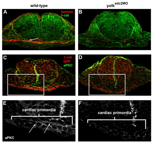
Basement membrane and epithelial cell polarity are disrupted in yolksdc2MO embryos. (A,B) Transverse sections of wild-type (A) and yolksdc2MO (B) embryos at 22 somites reveal laminin deposition (white arrow) between the cardiac and ventral foregut primordia in wild-type embryos (3/3) and reduced laminin in yolksdc2MO embryos (5/6). Laminin, red; β-catenin, green. (C-F) Transverse sections of Tg(cmlc2:GFP) embryos at 22 somites immunostained for the cell-junction protein aPKC (green) and cardiac primordia (red). Magnified views of cardiac precursors (marked by rectangles in C and D) reveal apicolateral localization of aPKC protein (arrows) in noninjected embryos (E) and diminished aPKC in yolksdc2MO embryos (F).
|