
Fig. 1
- ID
- ZDB-FIG-090904-64
- Publication
- Arrington et al., 2009 - Extra-embryonic syndecan 2 regulates organ primordia migration and fibrillogenesis throughout the zebrafish embryo
- Other Figures
- All Figure Page
- Back to All Figure Page
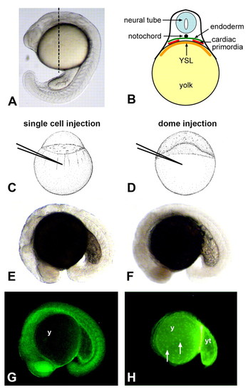
Testing the role of embryonic and nonembryonic cell lineages in cardiac and gut morphogenesis. (A) Lateral view of a zebrafish embryo at the 21-somite stage. Dashed line represents the approximate anterior-posterior location of cardiac primordia at this stage. (B) Diagram represents a transverse section at the level of the dashed line shown in A. Cardiac precursors migrate between the endoderm and the extra-embryonic yolk syncytial layer (YSL) towards the midline. Early gut primordia are positioned analogously but are located posterior to the cardiac primordia. (C,E,G) Morpholino injection into an embryo at the 1-cell stage (C), and light (E) and fluorescent (G) microscopy of a traditional morphant at the 18-somite stage (lateral view). In single-cell injections, fluorescent morpholino is enriched in embryonic tissues and is significantly reduced in the yolk (y). (D,F,H) Morpholino injection into an embryo at the dome stage (D), and light (F) and fluorescent (H) microscopy of a yolkMO embryo at the 18-somite stage (lateral view). In dome injections, fluorescent morpholino is restricted to the extra-embryonic yolk (y), posterior yolk tube (yt) and yolk syncytial nuclei (white arrows). Little, if any, fluorescence is detected within embryonic tissues. |