
Fig. 2
- ID
- ZDB-FIG-090904-32
- Publication
- Bollig et al., 2009 - A highly conserved retinoic acid responsive element controls wt1a expression in the zebrafish pronephros
- Other Figures
- All Figure Page
- Back to All Figure Page
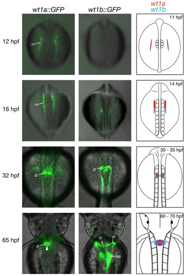
Reporter gene expression in transgenic zebrafish embryos recapitulates expression of wt1 paralogs. Transgenic wt1a::GFP (left) and wt1b::GFP (middle) embryos were imaged using a fluorescence microscope. Shown are overlays of dorsal transmission and fluorescence images at the indicated stages. Arrowhead in the bottom left panel marks fusion of the GFP signal at the midline. The schematic representation of wt1a and wt1b expression on the right is based on published data (Bollig et al., 2006; Drummond et al., 1998; Serluca and Fishman, 2001; Wingert et al., 2007) and is confined to expression in the pronephric region; wt1a expression is shown in red, wt1b expression in blue. ep, exocrine pancreas; gl, glomerulus; hpf, hours post fertilization; im, intermediate mesoderm; pt, pronephric tubule. |