
Fig. 7
- ID
- ZDB-FIG-090817-50
- Publication
- Curran et al., 2009 - Foxd3 Controls Melanophore Specification in the Zebrafish Neural Crest by Regulation of Mitf
- Other Figures
- All Figure Page
- Back to All Figure Page
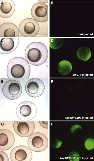
Foxd3 represses the mitfa promoter in zebrafish embryos. (A–H) One-cell mitfa:gfp transgenic zebrafish embryos microinjected with mRNA and imaged live at 6–7 hpf, shield stage 5x. (A, C, E, G) Brightfield images. (B, D, F, H) Green: live GFP expression from mitfa:gfp. (A, B) Un-injected embryos reveal no fluorescence at shield stage (observed in 64/64 embryos). (C, D) Embryos injected with sox10 mRNA produce robust, precocious mitfa:gfp expression at shield stage (observed in 62/64 embryos). (E, F) Co-injection of full-length foxd3 with sox10 mRNA prevents mitfa:gfp expression at shield stage (observed in 47/52 embryos). (G, H) Embryos co-injected with sox10 and DNA-binding mutant version of foxd3 mRNA display a return to robust, precocious mitfa:gfp expression at shield stage (observed in 43/49 embryos). |
Reprinted from Developmental Biology, 332(2), Curran, K., Raible, D.W., and Lister, J.A., Foxd3 Controls Melanophore Specification in the Zebrafish Neural Crest by Regulation of Mitf, 408-417, Copyright (2009) with permission from Elsevier. Full text @ Dev. Biol.