
Fig. 4
- ID
- ZDB-FIG-090804-58
- Publication
- Ng et al., 2009 - Zebrafish mutations in gart and paics identify crucial roles for de novo purine synthesis in vertebrate pigmentation and ocular development
- Other Figures
- All Figure Page
- Back to All Figure Page
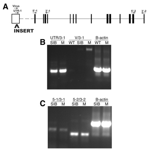
Molecular analysis of the garthi3526b locus. (A) The approximate position of the proviral insertion and RT-PCR primers mapped onto a schematic of the mutated gene (www.ensembl.org/Danio_rerio/). The proviral insertion is located in the predicted 5′ UTR. Amplification of the region between the UTR and exon 4 (B, UTR/3-1), the region between exons 1 and 4 (C, 5-1/3-1), or the region between exons 13 and 14 (C, 5-2/3-2) all indicate that the overall levels of gart transcripts are unaffected in the homozygous mutant embryos. Amplification from the 3′ end of the proviral insert to exon 4 (B, V/3-1) in homozygous gart mutants indicates that a portion of the proviral insert is transcribed and retained in the resulting mRNA. WT, wild-type AB; SIB, wild-type sibling mixture (+/+ and +/-); M, homozygous mutant. |