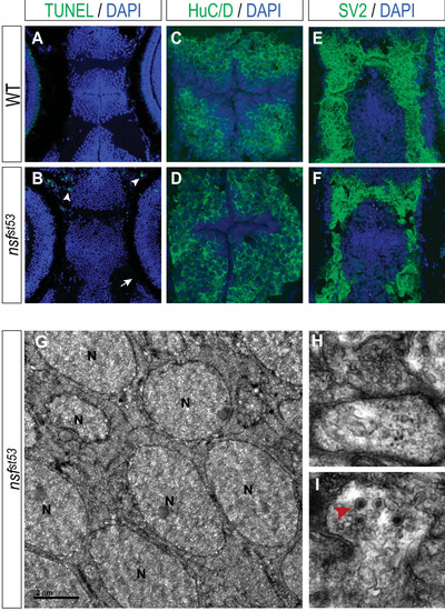
nsfst53 hypothalamus undergoes normal development and does not appear to initiate apoptosis. Apoptosis in (A) wild-type (WT) and (B) nsfst53 5 dpf zebrafish (12 μm horizontal sections) is shown. Cell death is not observed in the hypothalamus (B), but is observed in olfactory terminals (B, white arrowhead). Photoreceptor auto-fluorescence in wild-type zebrafish is lost in mutant (B, white arrow). DAPI staining of wild-type (A, C, E) and nsfst53 zebrafish (B, D, F) at 5 dpf is shown. Immunocytochemistry for (C, D) Hu family proteins and (E, F) SV2 are shown for wild-type and nsfst53 5 dpf zebrafish. All images are representative of at least 15 animals assayed. (G-I) Electron microscopy images from nsfst53 hypothalamic sections (n = 3). Cell bodies (G) and vesicle-filled synapse (H) resemble wild-type neurons (data not shown). Anatomical landmarks and (I) the presence of dark-cored secretory vesicles (red arrowhead) were used to confirm the location of the hypothalamus. N = nucleus. Note that the medially located cell bodies (Figure 4C, D, G) are framed on either side by lateral projections (Figure 4E, F, H) in both cryosection and EM images.
|
