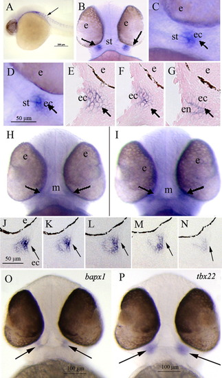
WISH analysis of zebrafish tbx22 expression in 38-42-hpf pharyngeal arch structures. B, H, I, O, P display ventral views, and all other panels present sagittal views. A: At 28 hpf, tbx22 is expressed in paraxial mesodermal tissues in a segmental pattern suggestive of vertebral pre-patterning (arrow). B, C: In 38-hpf embryos, tbx22 is first detectable as discrete, bilateral domains at either corner of the forming mouth (arrows). D: At 40 hpf, tbx22 is expressed in similar bilateral domains (arrow). E-G: Consecutive sagittal sections illustrate tbx22 expression underlying the bilaminar epithelium of the forming stomodeum, which bifurcates into dorsal and ventral elements (arrows). At 42 hpf, faint tbx22 expression is observed in tissues surrounding the mouth (H, arrows), and at each corner of the mouth opening (I, arrows), as shown in consecutive serial sections (J-N, arrows). O, P: At 50 hpf, bapx1 and tbx22 exhibit similar expression patterns in the developing jaw joint (arrows). e, eye; ec, ectoderm; m, mouth; s, stomodeum.
|
