
FIGURE
Fig. 2
- ID
- ZDB-FIG-090519-47
- Publication
- Lin et al., 1994 - lacZ expression in germline transgenic zebrafish can be detected in living embryos
- Other Figures
- All Figure Page
- Back to All Figure Page
Fig. 2
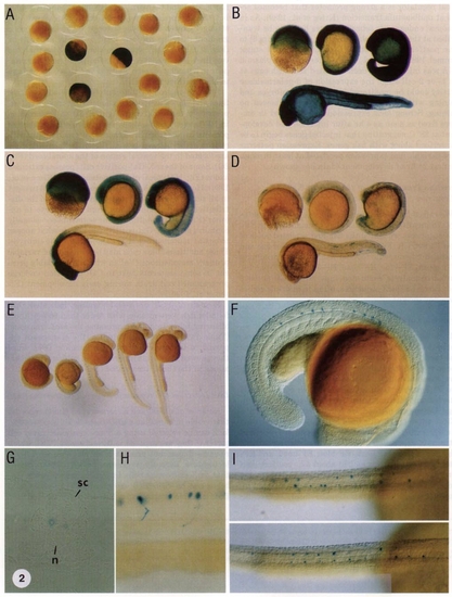
Expression of lacZ in F1 transgenic embryos. F1 embryos from line EF-head were fixed at the early gastrula and stained with X-Gal. Three embryos which express lacZ show readily discernible blue staining (A). Expression patterns of lines EF-blue (B), EF-head (C), EF-patchy (D), and EF-neuron (E) are shown at different developmental stages. Expression of lacZ in embryos from line EF-neuron is restricted to the CaP motoneurons in the spinal cord (F, G, and H). sc, spinal cord. n, notochord. The distribution of lacZ-expressing CaP cells is variable between embryos as shown from the dorsal view (I). |
Expression Data
Expression Detail
Antibody Labeling
Phenotype Data
Phenotype Detail
Acknowledgments
This image is the copyrighted work of the attributed author or publisher, and
ZFIN has permission only to display this image to its users.
Additional permissions should be obtained from the applicable author or publisher of the image.
Reprinted from Developmental Biology, 161, Lin, S., Yang, S., and Hopkins, N., lacZ expression in germline transgenic zebrafish can be detected in living embryos, 77-83, Copyright (1994) with permission from Elsevier. Full text @ Dev. Biol.