
Fig. 1
- ID
- ZDB-FIG-090519-46
- Publication
- Lin et al., 1994 - lacZ expression in germline transgenic zebrafish can be detected in living embryos
- Other Figures
- All Figure Page
- Back to All Figure Page
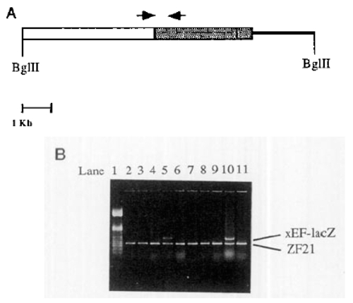
(A) Map of EF-lacZ. The EF1α enhancer-promoter is shown as a large open box, the lacZ gene is cross-hatched, and the plasmid vector pSP72 is shown as a line. The relative locations of the polymerase chain reaction (PCR) primers used to identify transgenic fish are indicated by arrows. (B) Identification of germline transgenic fish by PCR. Genomic DNA was extracted from a pool of embryos and was used in a PCR reaction with two pairs of primers. The first pair of primers is specific to the injected plasmid as shown in (A). The second pair of primers is specific to zebrafish homeobox gene, ZF-2.1 (Njolstad et al., 1988), generating a 475-bp product which is present in all the lines. Lane 1 is a 100-bp ladder. Lanes 2-9 are from pools of embryos from founder fish injected with EF-lacZ BglII fragment. Lane 10 is a positive control containing DNA from uninjected fish mixed with 20pg EF-lacZ plasmid. Lane 11 is a negative control containing DNA from uninjected fish. The founder fish in lane 5 (EF-50F) was detected as positive for the transgene. |
Reprinted from Developmental Biology, 161, Lin, S., Yang, S., and Hopkins, N., lacZ expression in germline transgenic zebrafish can be detected in living embryos, 77-83, Copyright (1994) with permission from Elsevier. Full text @ Dev. Biol.