
Fig. 3
- ID
- ZDB-FIG-090508-12
- Publication
- Riley et al., 1997 - A critical period of ear development controlled by distinct populations of ciliated cells in the zebrafish
- Other Figures
- All Figure Page
- Back to All Figure Page
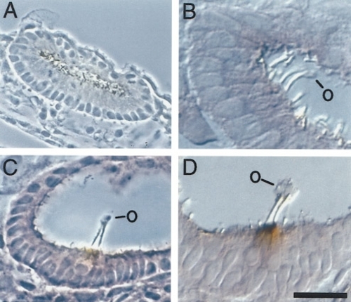
Sections of wild-type and mutant ears. Embryos were fixed and stained with peroxidase-conjugated antibodies to visualize acetylated tubulin and then sectioned. Specimens were lightly counterstained with eosin to help resolve cellular structure. (A) Phase-contrast image of the ear of a 19-h (20 somites) embryo showing numerous cilia. Although tethers cannot be clearly resolved at this time, all cells in the vicinity of the tether cells are columnar cells that fully span the otic epithelium, as indicated by the basal locations of their nuclei. (B) DIC image of the ear of a 20-h (22 somites) embryo showing two anterior tethers with apical otolith material (O). Tether cell bodies fully span the epithelium and show little or no staining for acetylated tubulin at this time. (C) Phase-contrast image of the ear of a 21.5-h (25 somites) embryo showing anterior otolith (O), tethers, and the overlapping bodies of two tether cells. The cytoplasm of tether cells counterstains less intensely than surrounding cells, a feature commonly observed in developing otic vesicles in both light and electron micrographs (Anniko, 1983; Noden and Van de Water, 1986; Hertwig and Schneider, 1986). The apical ends of tether cells stain positively for acetylated tubulin. (D) DIC image of the ear of a 21.5-h (25 somites) embryo showing anterior otolith (O), tethers, and tether cells. The distribution of acetylated tubulin at the apical ends of tether cells is clearly revealed in this lightly counterstained specimen. Anterior is to the right and dorsal is up in all panels. Scale bar, 20 (A), 15 (C), or 10 μm (B and D). |
Reprinted from Developmental Biology, 191(2), Riley, B.B., Zhu, C., Janetopoulos, C., and Aufderheide, K.J., A critical period of ear development controlled by distinct populations of ciliated cells in the zebrafish, 191-201, Copyright (1997) with permission from Elsevier. Full text @ Dev. Biol.