
Fig. 2
- ID
- ZDB-FIG-090508-11
- Publication
- Riley et al., 1997 - A critical period of ear development controlled by distinct populations of ciliated cells in the zebrafish
- Other Figures
- All Figure Page
- Back to All Figure Page
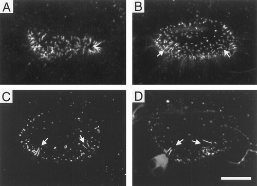
Distinct classes of cilia inside the otic vesicle. Embryos were fixed and incubated with primary antibodies directed against acetylated tubulin followed by fluorescent secondary antibodies. Stained embryos were then imaged by confocal microscopy to determine the distribution of cilia in the otic vesicle. (A) 19-h ear showing >100 cilia. (B) 24-h ear with two sets of presumed tethers (arrows) and numerous shorter cilia. (C) 27-h ear with two sets of tethers (arrows). Most shorter cilia have disappeared. (D) 30-h ear showing two sets of tethers. Anterior tethers are attached to hair cells the cell bodies of which stain intensely for acetylated tubulin. Posterior hair cells also stain positively but occur at a deeper (more medial) focal plane than is shown. Scale bar, 25 μm. |
Reprinted from Developmental Biology, 191(2), Riley, B.B., Zhu, C., Janetopoulos, C., and Aufderheide, K.J., A critical period of ear development controlled by distinct populations of ciliated cells in the zebrafish, 191-201, Copyright (1997) with permission from Elsevier. Full text @ Dev. Biol.