|
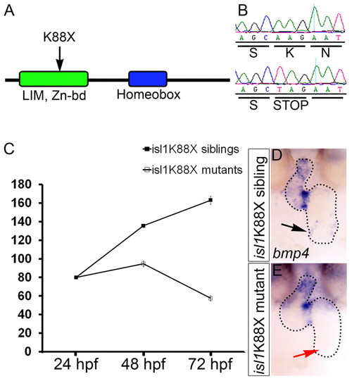
Cardiac defects of isl1K88X mutant embryos. (A) Cartoon of the Isl1 protein with the location of the premature stop codon (K88X) identified in the isl1 mutant. (B) DNA sequence of a homozygous wild-type sibling (top) and a homozygous isl1K88X mutant (bottom) at the site of the mutation (AAG>TAG) that results in a premature stop codon. (C) The number of heart beats per minute measured at 24, 48 and 72 hpf of wild-type sibling (black squares) and isl1 mutant (white squares) embryos. (D,E) In situ hybridization for bmp4 on 48 hpf wild-type sibling (D) and isl1 mutant (E) embryos. Black arrow (D) indicates bmp4 expression in the sinus venosus of a wild-type embryo; red arrow (E) indicates the inflow area of a representative isl1 mutant embryo lacking bmp4 expression. Embryos are shown as frontal-ventral views (OFT to the top).
|