
Fig. 4
- ID
- ZDB-FIG-090427-6
- Publication
- Wang et al., 2009 - Identification of wnt-responsive cells in the zebrafish hypothalamus
- Other Figures
- All Figure Page
- Back to All Figure Page
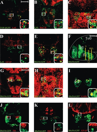
Immunohistochemical identification of Lef1- expressing cells in the 50 hpf embryonic hypothalamus. (A–E) Twelve-micron cryosections immunostained for the markers listed in each panel. Positions of cross sections for panels (A) and (C–E) are at the same level as Figure 3E, and panel (B) is at the same level as Figure 3G. Boxed region is shown at higher magnification in lower right corner, and circled cells are double labeled. Lef1 staining partially overlaps with TOP:dGFP, HuC/D, PCNA, and dlx5/6:gfp, but not with GFAP. (F) Lateral view of dlx5/6:gfp whole-mount brain, observed with a 100-μm confocal projection. (G–L) Twelve-micron cryosections immunostained for the markers listed in each panel. Positions of cross sections are indicated in panel (F). Boxed region is shown at higher magnification in lower right corner, and circled cells are double labeled. GFP staining partially overlaps with HuC/D, PCNA, and GABA, but not with TH, 5-HT, or GFAP. T, telencephalon; TeO, tectum opticum; DT, dorsal thalamus (thalamus); Hy, hypothalamus. Scale bars: (A, G) 50 μm; (F) 100 μm. |