
Fig. 2
- ID
- ZDB-FIG-090427-4
- Publication
- Wang et al., 2009 - Identification of wnt-responsive cells in the zebrafish hypothalamus
- Other Figures
- All Figure Page
- Back to All Figure Page
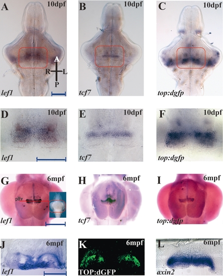
Expression of tcf genes and top:dgfp in the larval and adult zebrafish hypothalamus. (A–C) Ventral view of 10 dpf larval brains; orientation is indicated in panel (A). (D–F) Higher power images of the fields indicated in (A–C). top:dgfp Expression encompasses the combined domains of lef1 and tcf7. (G–I) Ventral views of 6mpf adult brains. (J) Cross section through adult hypothalamus at the level indicated in panel (G). (K) GFP antibody staining on an adult TOP:dGFP hypothalamus cross section at the same level as panel (J). (L) axin2 expression on an adult hypothalamus cross section at the same level as panel (J). lef1 and GFP/axin2 occupy different regions of the periventricular zone. pHy, periventricular hypothalamus. Scale bars: (A, D) 100 μm; (G) 500 μm; (J) 200 μm. |