
Fig. 8
- ID
- ZDB-FIG-090415-47
- Publication
- Fouquet et al., 1997 - Vessel patterning in the embryo of the zebrafish: guidance by notochord
- Other Figures
- All Figure Page
- Back to All Figure Page
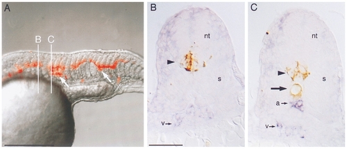
Mosaic analysis of flh embryos transplanted with wild-type cells. (A) Composite rhodamine epifluorescence (red) and Nomarski (gray) videomicrograph of the trunk of a 25 somite flh mutant embryo that was transplanted at the blastoderm stage. In this embryo wildtype cells contributed primarily to neural tube and notochord (arrows). B and C indicate planes of cross-sectioning in panels (B) and (C). (B) Cross section of flh embryo in plane B. Transplanted wild-type cells labeled with biotin– peroxidase (brown, arrowhead) contributed to ventral neural tube (nt). A single population of flk-expressing cells (blue, v) resides ventrally. The somites (s) are fused beneath the neural tube. (C) Cross section of flh embryo in plane C. Transplanted wild-type cells contributed both to ventral neural tube (arrowhead) and to notochord (arrow). There are two populations of flk-expressing cells, a dorsal one (a) just beneath the notochord and a ventral one (v). Somites (s) are not fused. Dorsal is up, anterior is to the left in (A). Bars, 250 μm (A), 50 μm (B, C). |
Reprinted from Developmental Biology, 183(1), Fouquet, B., Weinstein, B.M., Serluca, F.C., and Fishman, M.C., Vessel patterning in the embryo of the zebrafish: guidance by notochord, 37-48, Copyright (1997) with permission from Elsevier. Full text @ Dev. Biol.