
FIGURE
Fig. 5
- ID
- ZDB-FIG-090415-44
- Publication
- Fouquet et al., 1997 - Vessel patterning in the embryo of the zebrafish: guidance by notochord
- Other Figures
- All Figure Page
- Back to All Figure Page
Fig. 5
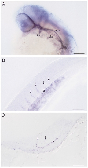
Whole mount RNA in situ hybridization with flk on 30 somite stage embryos (24 hpf) showing (A) a view of the head, (B) a lateral view of the trunk, and (C) a sagittal section of the posterior trunk and tail. (A) Clearly visible are the patent first aortic arch (aa) and left dorsal aorta (da). In contrast, the anterior cardinal vein (acv) does not yet possess a lumen. (B, C) In the trunk, intersomitic vessels (arrows) can be seen sprouting from the dorsal aorta (a). Dorsal is up and anterior is to the left. Bars, 100 μm (A), 50 μm (B, C). |
Expression Data
Expression Detail
Antibody Labeling
Phenotype Data
Phenotype Detail
Acknowledgments
This image is the copyrighted work of the attributed author or publisher, and
ZFIN has permission only to display this image to its users.
Additional permissions should be obtained from the applicable author or publisher of the image.
Reprinted from Developmental Biology, 183(1), Fouquet, B., Weinstein, B.M., Serluca, F.C., and Fishman, M.C., Vessel patterning in the embryo of the zebrafish: guidance by notochord, 37-48, Copyright (1997) with permission from Elsevier. Full text @ Dev. Biol.